Optical Coherence Tomography (OCT)
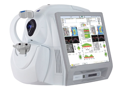
 OCT is a high resolution scanning device that analyzes the layers of the retina and optic nerve.
  It can be thought of as an MRI of the eye because of the fine detail it provides. It can detect subtle changes in the retina and optic nerve before they become detectable on an eye exam and before it begins to affect your vision. It is relatively new technology that has become an integral part of the diagnosis and treatment of glaucoma, macular degeneration and other retinal diseases.
Corneal Topography
Corneal topographers are imaging devices that map the shape and surface of the cornea which is the front layer of the eye. We use this instrument most often to monitor changes in the shape of the cornea due to contact lens wear, to assist in the fitting of Corneal Refractive Therapy (CRT) lenses and other specialty lenses, and as part of the screening process to determine whether or not a patient is a good candidate for LASIK. It is also useful in diagnosing corneal conditions such as Keratoconus and in examining patients who have unexplained reduced vision after any type of corneal surgery including LASIK.
Optomap Retinal Exam
 Checking the health of your retina is an important part of your comprehensive eye exam. The retina is the neurological tissue lining the inside of the eyeball. Without using dilating eye drops, only a small central area of the retina (about 30-40 degrees) can be seen.
Dilation of the pupils is the best way to view the entire retina and it allows the doctor a three dimensional view. The side effects of dilation are temporary blurriness and light sensitivity lasting about 3-4 hours. Some patients cannot be be dilated on the day of their exam because they are working or driving. Others cannot because of a sensitivity to the eye drops.
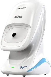
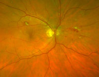
The Optomap is an FDA approved panoramic imaging device that provides a wide field view (about 200 degrees) of the retina without the need for dilating drops; hence, there are no blurry side effects. It is an excellent screening device, takes seconds to perform, and the results are available immediately.
It is also a way to document and monitor over time any changes within the eye. While it does not completely replace dilation, it can reduce the number of times your eyes will need to be dilated over the course of a lifetime and is a good alternative if you cannot be dilated the day of your exam. Please be aware that there is an additional fee of $30 for the Optomap scan.
Many retinal diseases are silent and do not have any symptoms until they are in very advanced stages. In addition, systemic diseases like high blood pressure and diabetes which can cause changes in the retina are often diagnosed for the first time during an eye exam. For more information, www.Optomap.com.

