| Blepharitis | Cataracts |
| Conjunctivitis | Diabetic Retinopathy |
| Keratoconus | Glaucoma |
| Macular Degeneration | Retinal Detachment |
Blepharitis
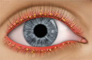 Blepharitis is an inflammation of the eyelids that causes red, itchy, irritated lids and a build up of dandruff-like debris on the lashes. It is a very common condition that affects people of all ages. There are two basic types of Blepharitis: Anterior and Posterior.
Anterior Blepharitis affects the front portion of the lids where the lashes grow out of. It is often caused by an overgrowth of bacteria (Staphyloccocus Blepharitis) or dandruff of the scalp and eyebrows (Seborrheic Blepharitis). Hormones, nutrition, general physical condition, and even stress may contribute to Seborrheic Blepharitis. Build-up of naturally occurring bacteria contribute to Staph Blepharitis.
Posterior Blepharitis affects the back portion of the lids which is in contact with the eyeball. Oil producing glands (meibomium glands) in this part of the lid work to keep our tears from evaporating too quickly. Posterior Blepharitis is caused by an abnormality in these glands which, over time, creates an environment in which bacteria will grow. People who have oily skin conditions such as Acne Rosacea often have Posterior Blepharitis.
SYMPTOMS
- Gritty burning sensation
- Dry eyes
- Itching and redness of the lid margins and/or eye itself
- Swollen red rimmed lids
- Crusting of the lids and lashes
- Loss of lashes or misdirected lashes
- Recurrent styes, hordeola or eye infections
- Excessive tearing
- Intermittent blurry or filmy vision
TREATMENT
Blepharitis is a chronic condition that requires treatment and on going maintenance which may consist of the following:
-
Warm compresses and lid massage to loosen up the crusting around the lashes. This also helps to make the oil producing glands in the lids function better.
-
Clean the lids using a mixture of either baby or tea tree oil shampoo and water. Gently scrubbing the lids and lashes will clean out the debris that has built up around the lashes and will mitigate the conditions in which bacteria tend to grow. Commercially prepared lid scrub products such as Sterilid or Ocusoft Lid Scrub pads are available.
-
Use an anti-dandruff shampoo when you wash your hair and scalp.
-
Use artificial tear drops to help with any dry eye symptoms.
-
Limit or temporarily discontinue eye make up as it adds to the debris around the lashes and makes cleaning the lids and lashes more difficult.
-
Temporarily discontinue wearing contact lenses during treatment as lens wear will likely not be comfortable when you have Blepharitis.
-
Some cases of Blepharitis, especially if the cause is primarily bacterial, may require low dose oral antibiotics or prescription eye drops and ointments.
Cataracts
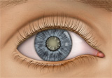 A cataract is a clouding of the lens inside the eye. The lens is made up mostly of water and protein arranged to let light through. When this water/protein balance is altered whether due to age, disease, or injury, light is blocked and the lens appears cloudy. Cataracts most commonly develop with age but can also be due to injury, diseases such as Diabetes, or as a side effect of certain medications. Some rarer forms of cataracts occur at birth.
SYMPTOMS
- Blurred vision
- Increase glare and sensitivity to bright lights
- Trouble seeing at night
- Colors look faded
TREATMENT
Cataracts, in and of themselves, are generally not harmful to the eyes and are easily detected during a routine eye exam. Mild cataracts often do not need treatment and simply need to be monitored. Vision generally can be improved with changes to one's eyeglass prescription with mild cataracts. If the cataract worsens and vision is impaired to a point where it can no longer be improved with glasses or it interferes with daily activities, surgery may be indicated. Surgery is done on an out-patient basis and involves a very small incision, through which the cloudy substance of the lens is removed and replaced with an artificial lens implant. Advancements in cataract surgery have made the recovery after surgery faster, easier, and more comfortable. Multifocal implants are now available that can correct both far and near vision so many patients after cataract surgery may no longer need glasses.
Conjunctivitis (Pink Eye)
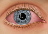 The conjunctiva is the transparent tissue that lines the inside of the eyelids and covers the white part of the eye. Conjunctivitis or "Pink Eye" is an inflammation of this tissue which causes the eye to appear red or pink. The most common causes of conjunctivitis are viruses, bacteria, and allergies.
GENERAL SYMPTOMS
- Mucus or watery discharge from one or both eyes
- Excessive tearing
- Itching, burning, or gritty sensation
- Swollen lids
- Whites of the eyes are pink
- Increase sensitivity to light
Viral Conjunctivitis is caused by viruses that are associated with the common cold. The individual will often have an accompanying cold or upper respiratory tract infection. It is spread by exposure to an infected person's coughing/sneezing or through the body's own mucus membranes as the lungs, throat, sinuses, nose and tear ducts are all connected. It is highly contagious so proper hygiene and precautions must be taken to prevent it from spreading. Individuals often have a very watery discharge and may be very light sensitive.
Bacterial Conjunctivitis is an infection caused by bacteria either from your own skin or respiratory system. It can be transmitted by contact with an infected person, dirty hands, or with contaminated eye make up or contact lenses. Patients often have green/yellow mucus discharge and mild itching. Bacterial infections can be very serious if the cornea (front layer of the eye) is affected as it can cause scarring and loss of vision if not treated properly.
Allergic Conjunctivitis most commonly occur in people who have seasonal allergies or allergies to specific substances such as cat hair, dander, or pollen. Itching is the main symptom along with tearing or mild mucus (usually white) discharge.
TREATMENT
The goal in treating conjunctivitis is to reduce the course of the infection or inflammation, to improve patient comfort, and to prevent it from spreading if it is viral or bacterial in nature.
Viral conjunctivitis, like the common cold, have to run their course which typically can take up to two weeks. There are no available drops to eradicate the virus and anitbiotics will not cure a viral infection. Cool compresses, artificial tear drops, and sometimes steroid eye drops can be prescribed to improve comfort. Some newer treatments include an iodine eye wash but there is no conclusive evidence that this shortens the course of the infection.
Bacterial conjunctivitis is typically treated with antibiotic eye drops or ointments, typically for 7-10 days. If the cornea is affected, treatment often needs to be more aggressive using stronger antibiotics dosed more frequently as bacterial corneal infections can lead to scarring and vision loss.
Allergic conjunctivitis is treated, first and foremost, by removing or avoiding the offending irritant. Cool compresses, artificial tear drops, antihistamine eye drops as well as over the counter allergy medications may be prescribed to improve comfort. In more severe cases, steroid eye drops may be needed to reduce the inflammation.
SELF CARE
Proper hygiene is the best way to prevent the spread of conjunctivitis:
- Wash your hands often and don't touch your eyes with your hands
- Don't share wash cloths or towels
- Discard and avoid using makeup which may have become contaminated
- Contact lens wear should be discontinued and lenses discarded
- Don't share contact lenses or cosmetics
- A child with pink eye should be kept from school for a few days
Diabetic Retinopathy
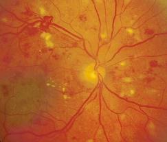 Diabetic Retinopathy is an eye disease associated with Diabetes. High levels of blood sugar can damage the blood vessels in the eye. This can lead to poor circulation in the eye and can cause small leaks in the blood vessels. The leaks can cause swelling of the retina which is the neurological tissue that lines the inside of the eye. Eventually new blood vessels may form to replace the damaged ones. These new blood vessels, however, are not normal and very fragile. They can burst, creating a hemorrhage in the eye, which can result in a permanent loss of vision.
SYMPTOMS
- Blurred or darkened vision
- Sudden loss of vision
- Diabetic changes in the eye often have no symptoms early on
RISK FACTORS AND MANAGEMENT
It is critical for all diabetic patients to have a thorough eye examination at least every year. When Diabetic Retinopathy is diagnosed early, treatment can often be implemented and is more effective in preserving vision. Diabetic patients also have a higher risk of developing cataracts at a younger age and of having glaucoma. Those at the greatest risk of developing retinopathy include:
- patients with poorly controlled Diabetes
- patients with juvenile or insulin dependent Diabetes
- patients who have had Diabetes for 10 years or more
- patients who also have high blood pressure or cardiovascular problems
If you have Diabetes, it is important to control your blood sugar levels in addition to any high blood pressure or other health problems you may have. Routine check ups with your physician are important. This will reduce your risk of getting diabetic eye disease and other complications involving the heart, kidneys, and other organs.
Keratoconus
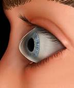 Keratoconus is a progressive vision disorder that occurs when the normally round cornea (the front part of the eye) becomes thin and irregular (cone) in shape. This prevents images from being focused correctly on the retina and causes distortion, often in the form of increasing amounts of astigmatism. In its earliest stages, Keratoconus causes slight blurring of vision and increased sensitivity to glare and light. These symptoms usually appear in the late teens or twenties. Keratoconus may progress for 10-20 years before slowing down. Both eyes are usually affected although unequally.
As Keratoconus progresses, the cornea bulges and becomes more and more cone shaped. Vision becomes blurrier, more distorted and frequent changes to one's eyeglass prescription are often needed. At some point, eyeglasses may longer provide adequate vision and specially designed contact lenses will be needed. These contact lenses are usually made of a rigid gas permeable material. Most cases of Keratoconus are managed with rigid contact lenses and frequent follow up visits.
In a small number of cases, the cornea will swell and cause a sudden decrease in vision. The swelling occurs when the strain of the protruding cornea causes a tiny break in the layers of the cornea. The swelling may last for weeks as the break heals and is gradually replaced by scar tissue. Eye drops can be prescribed for temporary relief. If the swelling recurs and scarring worsens, a corneal transplant may become necessary.
It is uncertain what causes Keratoconus but there is likely a genetic component. Eye rubbing as a child seems to be a common link in many cases. In individuals who are genetically predisposed, it is thought that eye rubbing sets off a process in the cornea that leads to thinning and an alteration in its shape.
A new treatment for halting the progression of Keratoconus, Collagen Cross Linking, is in the final stages of FDA approval. It involves the use of riboflavin eye drops and ultraviolet light to strengthen the structure of the cornea and thus halt the progession of the thinning.
Glaucoma
 The most common type of Glaucoma is caused by too much fluid pressure inside the eye. Fluid in the eye helps to nourish the eye and is constantly being produced and drained out of the eye through a very complex system of channels.
When either too much fluid is produced or its flow out of the eye is slowed or blocked, the internal eye pressure increases and can cause damage to the optic nerve. Damage to the optic nerve initially causes a loss of peripheral vision. Left untreated, this damage will eventually lead to total vision loss.
SYMPTOMS
Glaucoma is often called the "Silent Thief of Sight" because it has no symptoms until the disease is in advanced stages. Patients with advanced Glaucoma may have tunnel vision where the peripheral vision is very limited and only their central vision remains. Often, these patients can still read the small lines on the eye chart even though they have lost much of their overall vision.
RISK FACTORS AND MANAGEMENT
Risk factors for developing Open Angle Glaucoma include:
- Family history
- Age over 45 years
- African descent
- Near-sightedness
- Diabetes or Cardiovascular disease
- Smoking
- History of eye injury or severe blood loss due to surgery or trauma
- Long term use of certain medications such as steroids
Glaucoma testing is an important part of every routine eye exam and consists of checking internal eye pressure, peripheral vision, as well as evaluating the health of the optic nerves. Any suspicion of Glaucoma warrants further testing which includes more detailed peripheral vision testing and imaging of the nerve with sophisticated instrumentation.
The goal in treating Glaucoma is to lower the eye pressure in order to keep any vision loss from progressing. Treatment commonly consists of eyedrops and/ or laser therapy. Patients who do not respond well to these therapies or advanced cases of Glaucoma often need surgical intervention.
Macular Degeneration
 Macular degeneration (AMD) is generally an age related disease which affects the central portion of the retina known as the macula. The retina is the neurological tissue that lines the inside of the eye and the macula is the small central area of this tissue. The macula allows us to see the fine detail of whatever we look at directly. Macular Degeneration occurs when there is damage to this area.
WET VS DRY MACULAR DEGENERATION
AMD is often accompanied by the formation of yellow deposits called "drusen" under the macula which causes the retinal tissue to thin and atrophy. This is the most common form of the disease and is called "dry" AMD. Patients with the dry form often have mild to moderate blurred vision which may slowly worsen over time. In less common cases, abnormal blood vessels develop under the macula and leak fluid. This is called "wet" AMD and can cause severe vision loss.
RISK FACTORS
A number of uncontrollable factors contribute to AMD including age, sex (women more than men), eye color (light colored eyes seem to be at greater risk), farsightedness, and race (Caucasions are at higher risk). Controllable risk factors include smoking, high blood pressure, exposure to UV, and diet.
SYMPTOMS
The most common symptom is blurred vision. Wet AMD progresses much faster than the dry form and symptoms are more severe. Symptoms include blurred vision, distortion, or a dark spot in the central vision.
TREATMENT
Currently, there is no treatment for the dry form of the disease. Patients with the dry form need to be monitored routinely. Cessation of smoking, UV protection, improved diet with foods that are rich in certain vitamins such as lutein, zeaxanthin, and other antioxidants (leafy green vegetables and fruits) are preventative measures that patients can take. New treatments which involve the injection of drugs (Lucentis, Avastin) into the eye to halt or reverse the growth of new blood vessels under the macula are available for some patients with the wet form of the disease. The key, as with any disease, is early detection.
Retinal Detachment
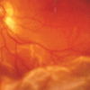 The neurological tissue that lines the inside of the eye is the retina. It functions to collect light and transmit images through the optic nerve to the brain which, in turn, tells us what we are seeing. This is essentially how the visual system works. When the retina separates from the walls of the eye, it is known as a Retinal Detachment.
SYMPTOMS
- A sudden loss of part or all of your vision
- A "shadow or curtain" that obscures part of your vision
- A sudden increase in "floaters," which look like small particles or fine threads
- Flashes of light
- No symptoms at all or a "blind spot" that is too small to notice
RISK FACTORS
- Eye injuries, tumors, and cataract surgery
- Near-sightedness
- Age
- Diabetics who have retinal disease (Diabetic Retinopathy)
- Family history of Retinal Detachment
TREATMENT
The key to successful treatment for Retinal Detachment is early detection. Detachments often start as a small tear in the retinal tissue. At this stage, there may be no symptoms and vision may not be affected and the torn retina is easily repaired. Left untreated, a retinal tear can progress and enlarge to a full detachment which can affect the vision and require more extensive surgery to repair. Treatments for retinal tears and detachments may involve the injection of either silicone or gas into the eye, laser, cryo therapy or implanting a silicone band over the eyeball in larger detachments. The longer the retina remains detached and untreated, the less likely vision can be recovered. That is why routinely dilating the eyes is important and any symptoms of flashes and floaters must be evaluated promptly.
